
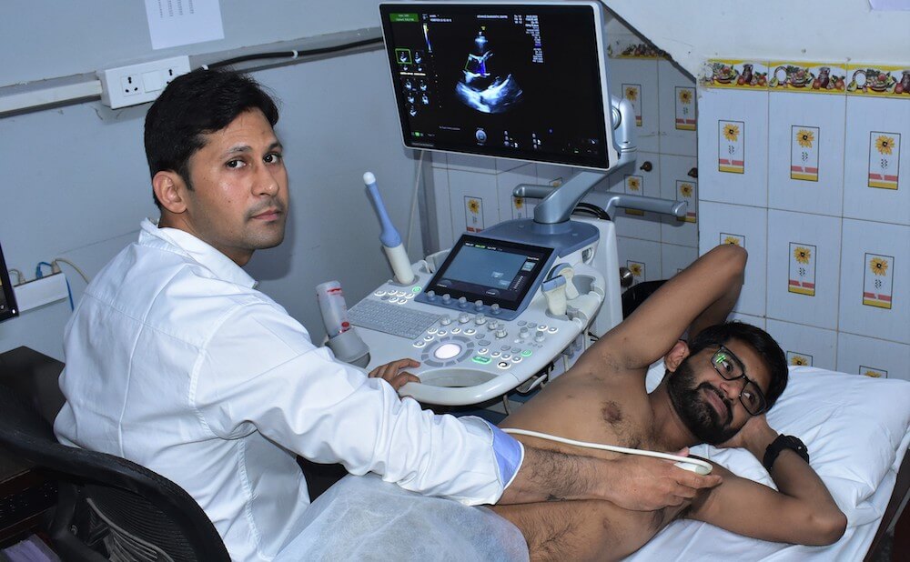
Echo Cardiography
A specialist called a cardiac sonographer performs your echo. Modern technology is used by our doctors. They have received training to perform tasks in a variety of settings, including catheterization laboratories and hospital rooms.
An EKG looks at your heart's electrical activity. It creates a graph rather than photos of your heart. Your heart rhythm and rate are depicted by the lines on this graph.
Why is an echocardiography performed during pregnancy?
A form of ultrasound test is foetal echocardiography. This test helps your doctor to get a better look at the anatomy and function of your unborn child’s heart.
A foetal echocardiography is not recommended for every pregnant woman. For the majority of women, a simple ultrasound will reveal the development of all four chambers of the baby’s heart. If prior tests were inconclusive or revealed an irregular heartbeat in the foetus, your OB-GYN may advise you to have this operation.
The findings of this test will help you and our doctors to arrange any post-delivery therapies for your baby. You can also seek assistance and counselling to help you make sound decisions for the remainder of your pregnancy.