
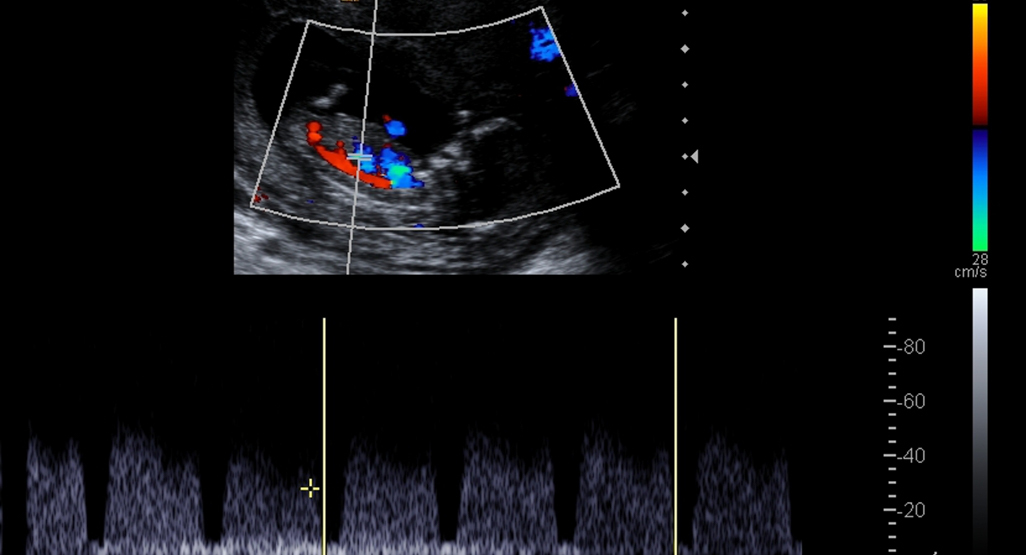
Color Doppler
The Color Doppler procedure typically involves the following steps:
Safety Considerations
Color Doppler imaging is generally considered safe and non-invasive. It does not involve ionizing radiation, making it suitable for repeated use and safe for pregnant women and children. Additionally, there are no known risks associated with the use of sound waves for imaging.
Applications of Color Doppler Imaging
Color Doppler imaging finds extensive use in various medical fields, including:
Cardiology: It helps diagnose heart conditions, such as valvular diseases and blood clots, by visualizing blood flow in the heart and major blood vessels.
Vascular Medicine: Color Doppler assesses blood flow in arteries and veins, detecting conditions like deep vein thrombosis (DVT) and peripheral artery disease (PAD).
Obstetrics: In prenatal care, it monitors blood flow in the umbilical cord and fetal vessels, ensuring the health of the developing baby.
Oncology: It aids in tumor characterization by assessing blood flow within tumors, which can help guide treatment decisions.
Benefits of Color Doppler Imaging
The advantages of Color Doppler imaging are manifold:
Real-Time Visualization: It provides immediate, real-time information about blood flow, enabling prompt diagnosis and treatment decisions.
Non-Invasive: Color Doppler is a non-invasive procedure that does not require the use of needles or contrast agents.
Versatility: It can be applied to various body areas and has a wide range of clinical applications, making it an invaluable tool in medical diagnostics.
Improved Patient Care: Color Doppler imaging enhances patient care by aiding in the early detection and monitoring of vascular and circulatory disorders.