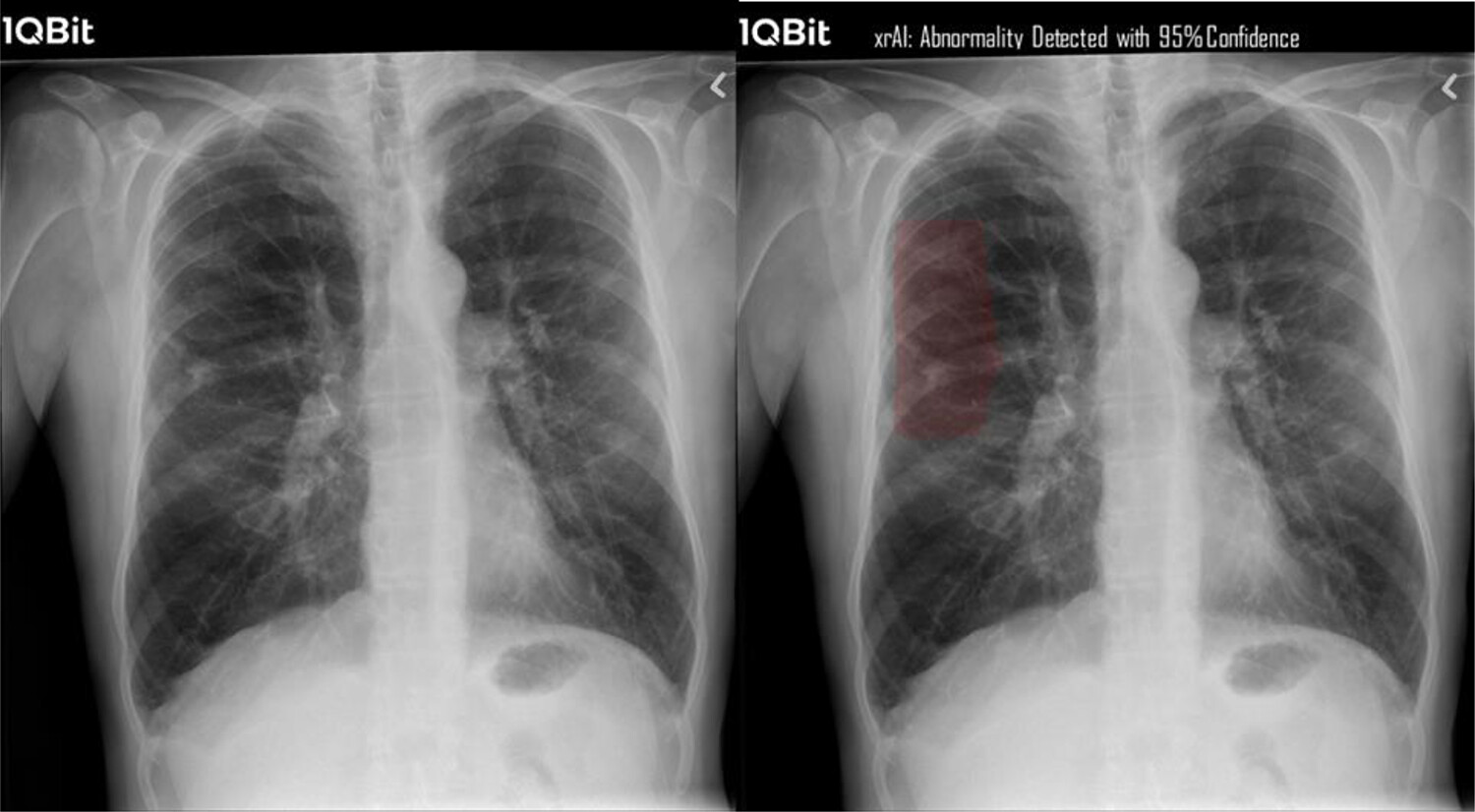
X-Ray

X-ray imaging, also known as radiography, is a widely used medical imaging technique that allows healthcare providers to visualize the internal structures of the body, such as bones, organs, and tissues, using X-rays. Here’s an overview of X-ray imaging:
- Principle of X-ray Imaging: X-ray imaging works on the principle of differential absorption of X-rays by different tissues in the body. When an X-ray beam passes through the body, it is absorbed by dense tissues such as bones, resulting in decreased transmission of X-rays to the detector. Soft tissues, such as muscles and organs, allow more X-rays to pass through and reach the detector.
- X-ray Machine: An X-ray machine consists of a tube that emits X-rays and a detector (film or digital sensor) that captures the X-ray images. The patient is positioned between the X-ray source and the detector, and the X-ray beam is directed towards the area of interest.
- Diagnostic Applications: X-ray imaging is used to diagnose a wide range of medical conditions and injuries, including fractures, dislocations, pneumonia, lung diseases, gastrointestinal obstructions, dental problems, and foreign objects in the body. X-rays are also used in interventional procedures, such as fluoroscopy-guided injections and angiography.
Types of X-ray Examinations:
- Plain X-rays (Radiographs): Plain X-rays produce 2D images of the body’s internal structures. They are commonly used to assess bone fractures, joint abnormalities, and chest conditions.
- Fluoroscopy: Fluoroscopy is a real-time X-ray imaging technique used to visualize moving structures, such as the digestive system (barium swallow or upper GI series), blood vessels (angiography), and the urinary tract (voiding cystourethrogram).
- Computed Tomography (CT): CT scans combine X-ray images taken from multiple angles to create detailed cross-sectional images of the body. CT scans are used to evaluate internal organs, detect tumors, assess trauma, and guide surgical planning.
- Mammography: Mammography is a specialized X-ray examination used for breast cancer screening and diagnosis. It helps detect breast abnormalities, such as masses or calcifications, at an early stage.
- Safety Considerations: While X-ray imaging is generally safe and painless, exposure to ionizing radiation carries a small risk of potential harm, especially with repeated or high-dose exposures. To minimize radiation exposure, healthcare providers use the lowest possible radiation dose necessary to obtain diagnostic images and employ safety measures such as lead shielding and collimation.
- Digital Radiography: Digital radiography has largely replaced traditional film-based X-ray imaging. Digital X-ray systems use digital detectors to capture and process X-ray images, offering advantages such as shorter examination times, improved image quality, and easier image storage and retrieval.
In summary, X-ray imaging is a valuable diagnostic tool used by healthcare providers to visualize and assess a wide range of medical conditions. With ongoing advancements in technology and imaging techniques, X-ray imaging continues to play a crucial role in medical diagnosis, treatment planning, and patient care.
